Characterization of Metabolites Production by Lactobacillus Gasseri ATCC 33323 and Antioxidant Activity - Juniper Publishers
Nutrition and Food Science International Journal - Juniper Publishers

Abstract
Lactic acid bacteria (LAB) play an important role as natural food preservatives. However, the characterization of the variety of their metabolites is limited. The aim of this study was to determine specific metabolites produced by Lactobacillus gasseri ATCC 33323 by an optimized liquid chromatography with an ultraviolet/diode detection (HPLC-UV/DAD) method and to investigate their potential antimicrobial activity against specific food pathogens (Salmonella enterica ATCC14028; Staphylococcus aureus ATCC 29213; Escherichia coli ATCC 25922 and Klebsiella pneumoniae ATCC 700603). At the same time, the possible antioxidant activity of metabolites of L. gasseri was tested using the free radical DDPH• (a, a-Diphenyl-β-Picrylhydrazyl). Based on the results of this study, the major metabolites detected in L. gasseri were 6.0 ppm OH-PLA, 3.73 ppm 1,2-dihydroxybenzene, 2.31 ppm benzoic acid in 5 days. This study provides a different alternative approach to the others involved in the antimicrobial activity of food fermented by microorganisms. These molecules can be used as antimicrobial ingredients in the food industry instead of conventional chemical preservative.
Keywords: LAB; Metabolites; HPLC-DAD; Antioxidant activity
Abbreviations: LAB: Lactic acid bacteria; GRAS: Generally Recognized as Safe; EFSA: The European Food Safety Authority; FAO: The Food and Agricultural Organization of the United States; LLE: Liquid liquid extraction; CFSs: cell-free supernatant; RT: room temperature
Introduction
In recent years the addition of chemical preservatives has fallen into displeasure with consumers who are on the lookout for high quality, less heavily preserved, less severely processed, more natural (free of artificial additives) and safer food [1-3]. On the other hand, unprocessed foods can abet dangerous pathogens which can breed under refrigeration and without oxygen. A recourse to this dilemma is bio preservation, or more specifically the use of antimicrobial metabolites of fermentative microorganisms, such as lactic acid bacteria (LAB) [4]. The definition of bio preservation is the extension of shelf life and enhanced safety of food using natural and controlled microbiota and/or antimicrobial compounds [5,6]. Great efforts have been made to use the action of probiotic microorganisms and their antimicrobial products as a strategy for food control and bio preservation [7, 8].
Several microorganisms, especially LAB, with antimicrobial characteristics, have been commonly associated with food. LAB are a group of Gram-positive organisms, some of which have been classified as “Generally Recognized as Safe (GRAS)” by The European Food Safety Authority (EFSA) and The Food and Agricultural Organization of the United States (FAO) [9-10]. The use of LAB strains as probiotic and as bioprotective culture in fermented products has also been widely investigated [11-15].
It is therefore understood that LAB are extensively used in the fermentation of a variety of food products and are known for their curative and preservative effects [16]. This inhibitory effect of bacteria against pathogens is mainly due to the production of lactic acid and metabolites (e.g., bacteriocins, organic acids, diacetyl etc.). Metabolites are intermediate or end products of metabolism. They have a variety of functions, such as lowering pH with acid production exerting antimicrobial action extending shelf life with enhanced [17-20]. In addition to this, metabolites generated from LAB are known to reduce antimicrobial resistance [21]. These compounds can be effective against both Gram negative and positive bacteria, increasing their importance for natural preservation [22]. However, the characterization of the produced metabolites has still not been fully investigated. One of these cases is the identification of Lactobacillus gasseri’s metabolites.
Lactobacillus gasseri was discovered in the early 1970s by a group of strains, which up to that time belonged to the species Lactobacillus acidophilus. L. gasseri cannot be distinguished from L. acidophilus with classic phenotypic characteristics. However, L. gasseri was found to differ from L. acidophilus in the electrophoretic motility of its LDH (D- and L- lactate dehydrogenases) and in its cell wall composition [23]. LDH is the catalyst that helps convert lactic acid to pyruvate in the glycolysis cycle. One of these groups was named L. gasseri. Lactobacillus gasseri sp. nov., a new species of the subgenus Thermobacterium. Zbl Bakteriol Mikrobiol Hygiene I Abt Originale C1: 75-78) [24, 25].
The aim of this study was to determine specific metabolites produced by Lactobacillus gasseri and to investigate their potential antimicrobial activity against common food pathogens. Considering that LAB exhibit similar effects in vitro and the fact that their metabolites may target and play a role in the competitive exclusion of pathogens, the present study aimed to screen bacteria that produce numerous antimicrobial peptides and acids that may be effective bio preservatives for food products.
Materials and Methods
Bacterial Strain and Culture Conditions
Lactobacillus gasseri ATCC 33323 was grown in MRS broth Lactobacillus gasseri ATCC 33323 was grown in MRS broth (De Man Rogosa Sharp) and incubated at 37 °C for 48 hours under anaerobic conditions using AnaeroPack-Anaero (Mitsubishi Gas Chemical Company, Inc., Tokyo, Japan). The stock cultures were kept at -80°C in MRS broth containing 20% (v/v) sterile glycerol.
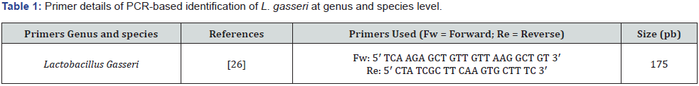
Genomic DNA Extraction: DNA was directly extracted from samples using an automatic extractor with the Nucleic Acid Extraction Kit, (ZYBIO Company), following the protocol recommended by the supplier. The purity and the quantity of the extracted DNA was evaluated spectrophotometrically by calculating OD260/OD280 (Epoch spectrophotometer, BioTek, Winooski, VT, USA). PCR was performed in 50μL final volume solution using the Master Mix (Hot Start Promega). The amplification was conducted by a thermal cycler (Veriti™ 96-Well Fast Thermal Cycler from Applied Biosystems, Thermo Scientific, US), as follows: an initial denaturation at 95°C for 3 min; followed by 35 cycles with the following step-cycle profile: denaturation at 95°C for 30s; annealing at 54°C for 60s ; extension at 72°C for 60s, and a final extension at 72°C for 10 min. PCR products were separated in 2% agarose gels, stained with ethidium bromide (0.5μg/mL), and documented under UV illumination using MiniBIS Pro device (DNR Bio- Imaging Systems Ltd., Neve Yamin, Israel) (table 1).
Pathogenic Microorganisms
The pathogenic microbial species Salmonella enterica ATCC14028, Staphylococcus aureus ATCC 29213, Escherichia coli ATCC 25922 and Klebsiella pneumoniae ATCC 700603 were used to study the antimicrobial activity of the metabolites of the selected lactic acid bacteria.
Preparation of Standards
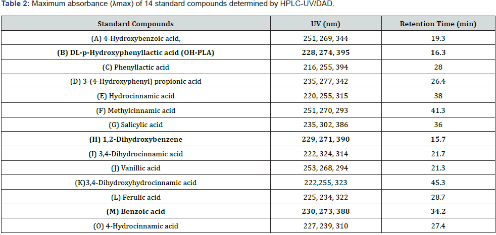
The standard solutions (table 2) were prepared at known concentrations (100ppm) in 90/10 (H2O / Methanol). The calibration curve standards were prepared at 1, 5, 10, 15 and 20 ppm and stored in vials (1.5mL) supplemented with 90/10 (H2O / Methanol). The selection of the standard compounds was based on literature [27].
Extraction Procedures
The Lactobacillus gasseri ATCC 33323 was grown in MRS broth at 37°C for 48 hours under anaerobic conditions. The broth was centrifuged at 13.000g for 7 minutes and the supernatants was filtered (filter pore size 0.22μm) to prepare cell-free supernatants (CFSs). A non-inoculated MRS broth medium (no bacteria added) was used as a blank matrix (negative control).
Liquid Liquid Extraction (LLE)
Ten mL CFSs were added to 10mL ethyl acetate, 1g NaCl and 4g Na2SO4 in 50mL conical tubes and centrifuged at 4,000g for 10 min. Subsequently, 5mL of the organic phase was collected by adding 100μl of dimethyl sulfoxide (DMSO) and let dry in a rotary evaporator. The dry phase was reconstituted (90% water and 10% methanol). The sample saw filtered (0.22μm) and aliquoted in two different vials. The 14 standards of known concentration were added in the first aliquot as internal controls to identify exactly the retention time and the wavelength of each peak. Both aliquots (with and without the internal controls, respectively) were analyzed by HPLC-UV/DAD. The optimum separation conditions are described below. A blank MRS broth was treated accordingly [27].
Ten mL CFSs were added to 10mL ethyl acetate, 1g NaCl and 4g Na2SO4 in 50mL conical tubes and centrifuged at 4,000g for 10 min. Subsequently, 5mL of the organic phase was collected by adding 100μl of dimethyl sulfoxide (DMSO) and let dry in a rotary evaporator. The dry phase was reconstituted (90% water and 10% methanol). The sample saw filtered (0.22μm) and aliquoted in two different vials. The 14 standards of known concentration were added in the first aliquot as internal controls to identify exactly the retention time and the wavelength of each peak. Both aliquots (with and without the internal controls, respectively) were analyzed by HPLC-UV/DAD. The optimum separation conditions are described below. A blank MRS broth was treated accordingly [27].
Instrumentation and Analytical Conditions
Chromatographic Conditions (HPLC-UV/DAD): A Hitachi LaChrom Elite HPLC system with diode array detector (L-2455) achieved separation of standards compounds on a SVEA C18 Gold column (150x4.6 mm 5μm, Sweden). The column was maintained at 30°C. The injection volume was 20μl and λmax=280nm however a λ range of between 210-325nm. Elution was performed using a gradient flow from water containing 0.1% formic acid (solvent A) and methanol containing 0.1% formic acid (solvent B). Initial conditions were optimized (0 min - 10% (B); 10 min - 30% (B); 20 min - 40% (B); 25 min - 40% (B), 40 min - 100% (B); 45 min – 100% (B); 50 min – 90% (B); 55 min – 10% (B)) at a flow rate of 0.500mL / min-1 [27]
Precision of standard calibration curves: Linear calibration curves were determined by generating a standard curve, in which serial 2-fold dilutions of standards were analyzed. The standard curve was generated by plotting the absorbance of each dilution against the known concentrations of standards. The resulting slope showed a linear relationship over 5 orders of magnitude, ranging from 1.25 to 20mg/mL with a correlation coefficient R2 >0.99. The detection rate was 100 % for up to 1mg/mL figure 1.
Antimicrobial Activity Assay
Four standardized ATCC strains from laboratory stock cultures were used in the evaluation of the antimicrobial activity of the CFSs, i.e., Salmonella enterica ATCC14028, Staphylococcus aureus ATCC 29213, Escherichia coli ATCC 25922 and Klebsiella pneumoniae ATCC 700603. Testing media (Selective Chromoagar for each pathogen) were used to evaluate the antimicrobial activity. The antimicrobial activity of CFSs filtrate was determined against the pathogen’s organisms. The CFSs incubated for 5 days at 37°C. Fifty microliters of the incubated CFSs were removed on different days to find the best antimicrobial activity. One hundred microliters of microbial culture of an approximate inoculums size of 1.0×10⁸ CFU/mL was added to all wells. Fifty microliters of the CFSs, on different days of incubation, were loaded into the microtiter plate containing each of the tested microbial strains. The plate was then incubated at 37°C for 24h. The lowest effective dose of each selected supernatant, which still significantly inhibited the growth of target pathogens, was determined as described in table 3,4. The antimicrobial activity was confirmed using selective agar plates for each pathogen, as shown. All measurements were performed in triplicate [27].
Assay Antioxidant Activity of LAB Strain in vitro
Scavenging of a, a-Diphenyl-β-Picrylhydrazyl (DDPH) Free Radical
Scanning results on LAB DPPH were measured by a modification of the method used by Kim et al. and Lee et al. [28,29]. The DDPH• is a violet-colored stable free radical, which is reduced to 2,2-diphenyl-1-picrylhydrazine (pale yellow), by reacting with an antioxidant [30]. The DDPH• solution of 6x10- 5 M was prepared in methanol. 1.0mL of cell-free supernatants (CFSs) were added to 2.5mL of ethanolic DPPH radical solution (Asample). The sample was incubated at room temperature (RT) in the dark for 30 minutes after mixed vigorously. The absorbance of the DDPH radical without CFSs was also measured (Acontrol). Absorbance of the supernatant was measured in triplicate at 517nm. Total antioxidant activity is expressed in μg/ml gallic acid. The results are expressed as the amount of antioxidant needed to cause a 50% reduction in DDPH absorption (IC50). The radical scavenging activity was computed using the following equation:
Radical scavenging activity (%) = [ (Acontrol – Asample) / Acontrol] × 100%
where Asample = absorbance of sample; Acontrol = absorbance of control
Results and Discussion
Historically lactic acid bacteria (LAB) have been used in many industrial fermentation processes. More recent applications including the use of LAB as probiotics [30-32] have significantly increased industrial interest. The genus Lactobacillus is one of the most important genera of lactic acid bacteria and the great technological interest of lactobacilli is perceived by the high number of researches that have been done for their physiology, the existence of probiotic properties and the production of antimicrobial compounds.
The extracted DNA collected was checked for its concentration and purity by photometry. All samples had a concentration of 80-120ng/μl and a purity of 1.5-1.9ng/μl. Electrophoresis on 2% agarose gel was performed to verify the method. A PCR of a known molecular weight of 100bp (ladder) was added during the electrophoresis of the PCR products to calculate the molecular size of the fragment. The amplified segments were 175bp in size. The figure 1 shows a typical result of a PCR amplification of the portion of the gene studied.
LAB have an important antimicrobial function due to their production of certain metabolites, such as organic acids [33-37]. The identification of standards, the wavelengths and the retention time of each compound are shown in table 2. The response intensity and retention time of the chromatographic peaks from the three analyses almost overlap, which shows limited variations due to instrument errors. The chromatographic method was optimized for the quantitative analysis of 14 compounds in LAB strains. within a run period of 60min.
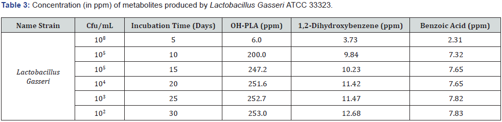
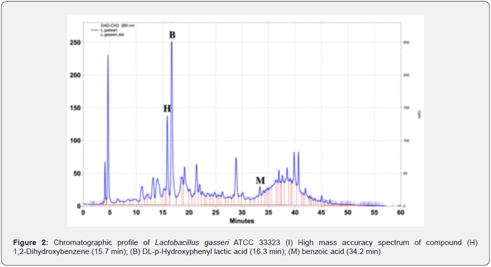
The main metabolites that were detected in Lactobacillus gasseri ATCC 33323 presented in figure 2 and table 3. The concentration of each metabolite was determined 6.0ppm of OHPLA, 3.73 ppm of 1,2-Dihydroxybenzene, 2.31ppm of benzoic acid in 5 days. In the present study, the concentration of metabolites increased until the 15th day. Afterward, the metabolites’ concentrations followed a stable pattern [26]. It was observed that the metabolites produced by Lactobacillus gasseri ATCC 33323 in the broth medium remained the same over a period of 30 days while the concentrations increased in the broth (table 3). Thus, metabolites could be identified from the first day of incubation of the microorganism. None of these compounds was detected in the blank MRS broth. The analysis was implemented for 30 days period. The concentration of each metabolite was determined at 5 days interval, as shown in table 3.
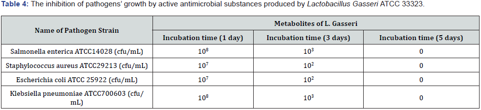
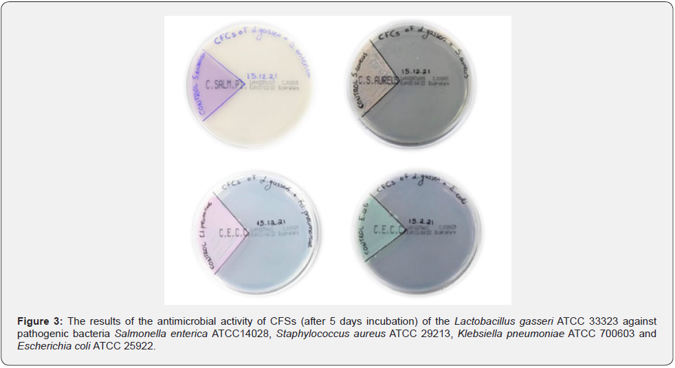
The present study investigated the antimicrobial activity of the obtained CFSs after 1st, 3rd, 5th days incubation of Lactobacillus gasseri ATCC 33323 against pathogenic microbial species (Salmonella enterica ATCC14028; Staphylococcus aureus ATCC 29213; Escherichia coli ATCC 25922; Klebsiella pneumoniae ATCC 700603). The inhibition of the active antimicrobials produced by L. gasseri after incubation at 37°C against these pathogens,shown in table 4 and figure 3. All CFSs obtained from Lactobacillus gasseri ATCC 33323 showed significant inhibitory effects on the tested Gram negative and positive pathogens. Isolated LAB has successfully inhibited the growth of tested pathogens, suggesting that the addition of this LAB strain to commercial foods may provide effective protection against infections caused by specific pathogens.
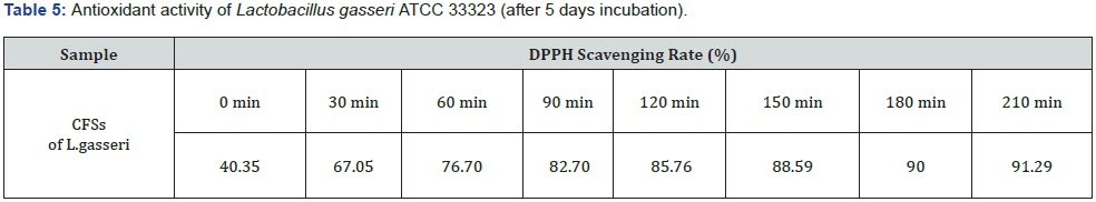
DPPH free radical is a stable radical with an unpaired valence electron at one atom of nitrogen bridge. Scavenging of DPPH radical is the basis of the popular DPPH antioxidant assay. Oxidative stress results from a disequilibrium between oxidant and antioxidant actions. In our study, in vitro antioxidant activity of CFS from L. gasseri was measured through the DPPH radical scavenging method (table 5). The absorbance of different CFSs of L. gasseri concentrations was measured at different times (0min, 30min, 60min, 90min ,120min, 150min, 180min, 210min). The lowest value corresponds to the best antioxidant activity of the sample at 210minutes (91.29%).
At the same time, the antioxidant activity of the three standards that were detected and quantified in CFS from L. gasseri was studied. After 90min in the darkness, the scavenging rate of the mix of three standards is decreasing. In this time, the highest antioxidant is achieved (74.82%). On the other hand, the highest antioxidant activity of the sample was measured at 210min (91.29%) (table 6). Over this period the percentage is reduced. In conclusion, that there are other metabolites in the CFS that help in better antioxidant activity.
Conclusions
LAB are the most used microorganisms for the fermentation and preservation of foods. The increasing societal demand for less processed food products, while conserving those products’ quality, safety, and shelf-life has raised the question of chemical preservative replacement. The present study investigated the antimicrobial activity of CFS obtained by Lactobacillus gasseri ATCC 33323 from the 1st - 30th day of incubation against four pathogenic foods (Salmonella enterica ATCC14028, Staphylococcus aureus ATCC 29213, Klebsiella pneumoniae ATCC 700603 and Escherichia coli ATCC 25922).
Complete inhibition of all pathogens was achieved after the 5th day of incubation at 37°C by Lactobacillus gasseri ATCC 33323. In this context, L. gasseri and its metabolites are alternatives of interest for use in food as bioprotective tools to combat microbial pathogens and to meet consumer demands. However, only 3 natural antimicrobials were detected and quantified in the CFS obtained. From the resulting spectra (Figure 2) several peaks that have not been previously identified were observed. Optimization of analytical assays can provide powerful tools for the identification of currently unknown compounds produced by LAB strains, which are involved in antimicrobial mechanisms against various food pathogens and spoilage microorganisms. This may include validating the proposed analytical method in real food systems and controlling antibacterial activity not only in vaccinated bacteria but also in the natural microflora of different food matrices.
To Know More About Nutrition and Food Science International Journal
Please click on: https://juniperpublishers.com/nfsij/index.php
For more Open Access Journals in Juniper Publishers
please click on: https://juniperpublishers.com/index.php
Comments
Post a Comment